
Journal of Advanced Biological Sciences
ISSN Print: N/A
ISSN Online: 3134-8823
About: Journal of Advanced Biological Sciences (JABS) is a peer-reviewed, open-access journal that aims to publish cutting-edge research and advancements in all areas of biological sciences. The journal serves as a platform for researchers, academicians, and professionals to contribute their scientific knowledge and foster innovation in biology-related fields.
Journal of Advanced Biological Sciences | Year 2024 | Volume 1 | Issue 1 | Pages 16-21
Synergistic and Antagonistic Effects of Cnicus Benedictus Floral Extract with Vinblastine on Cervical Cancer Cell Proliferation
Sadeq Jaafer Al-Tameemi 1* , I. Marwan Al-Zuhairi2 and M. Diyar Jalil31,2Department of Pharmacy, Bilad Alrafidain University College, Diyala, 32001, Iraq.
3Department of Pharmacy, Al-Yarmok University College, Diyala, Iraq
View PDF Download XML Download DOI XML DOI: 10.66590/jabs2024010103
Abstract
Background: This study aimed to evaluate the antiproliferative effect of Cnicus benedictus floral extract on cervical cancer cells under in vitro conditions. Additionally, the study investigated the potential synergistic or antagonistic interactions between the plant extract and Vinblastine. Methods: The HeLa cell line was used to assess the cytotoxic effects of Cnicus benedictus floral extract, vinblastine and their combination. The tested concentrations ranged from 1 to 10,000 µg/mL, evaluated at 24 and 72 hours of incubation. Drug interaction analysis was performed using the Compusyn software to calculate the Combination Index (CI) and Dose Reduction Index (DRI). Results: The findings demonstrated that the plant extract significantly inhibited cervical cancer cell proliferation in a concentration- and time-dependent manner. A similar trend was observed for the combination treatment. In contrast, the cytotoxic effect of vinblastine was primarily time-dependent. Drug interaction analysis revealed variable effects depending on concentration. At lower concentrations, the combination exhibited synergistic effects, particularly at 24 hours of incubation. However, moderate and higher concentrations showed antagonistic interactions. The Dose Reduction Index indicated a favorable reduction in vinblastine concentration at 24 hours when used in combination with the plant extract. Conversely, at 72 hours, both vinblastine and most extract concentrations demonstrated an unfavorable dose reduction profile. Conclusion: The alcoholic extract of Cnicus benedictus exhibited significant antiproliferative activity against cervical cancer cells, comparable to vinblastine. The combination treatment showed dual interaction patterns, with synergism at lower concentrations and antagonism at moderate to higher concentrations. Furthermore, the results suggest that the plant extract may exert both cell cycle–specific and non-specific cytotoxic effects, whereas vinblastine predominantly acts through cell cycle–specific mechanisms.
INTRODUCTION
Neoplastic diseases remain the greatest reason for mortality globally, rating second after cardiovascular diseases [1]. Globally, cancer accounts for one out of every eight fatalities. The number of deaths provoked by cancer is higher than the combined number of deaths caused by malaria, HIV and tuberculosis diseases [2]. Cervical cancer is a prevailing malignant illness that mostly involves women, with a global occurrence of almost 500,000 new cases each year. It rates second in terms of commonness after breast cancer [3].
Chemotherapy is often considered the primary treatment for cervical cancer. In the earlier stages of the disease, a combination of surgery or radiation therapy with chemotherapy is utilized. During advanced stages, the primary treatment often concerns a combination of radiation and chemotherapy. Chemotherapy is often utilized as a treatment for progressive cervical cancer [4]. Chemotherapy medications selectively eradicate cancer cells while simultaneously causing injury to some normal cells that experience rapid division, resulting in specific adverse outcomes. The effects vary relying on the specific medications and the dosage Utilized [5].
The common side effects of chemotherapy include vincristine-possess neurotoxicity and methotrexate-possess nephrotoxicity [6].
Cnicus Benedictus L is a plant species that has an antineoplastic effect. It is predominantly located in the northern region of Iraq and blossoms throughout the winter and spring seasons. The antineoplastic and antioxidant capacities of Cnicus Benedictus L are remarkable and the geographical zone and growing time affect their efficiency [7-9].
Multiple studies were conducted to assess the anticancer effect of Blessed Thistle. One of these studies examined the ethanol-based extract obtained from the flowers of Cnicus Benedictus L. It was found that this extract can effectively destroy mammary adenocarcinoma cancer cells (AMN-3). The effectiveness of the extract depends on each dosage and course of exposure [10]. On the other hand, several studies were established to evaluate the effectiveness of mixing Traditional chemotherapy with various natural substances in treating cancer; a particular study precisely assessed the inhibitory effect of vinblastine, in conjunction with laetrile, on the growth of cervical cancer cells. The study's results exhibited a synergistic interaction between the components in the combination, offering a powerful anticancer effect [11-13].
Several investigations have been carried out on this issue, demonstrating a shortage of adequate data on the Antiproliferative ability of the Cnicus Benedictus flower on cervical cancer cell lines, as well as its possibility when mixed with other Traditional chemotherapy treatments. The purpose of our study was to assess the cytotoxic impact of the floral extract of Cnicus Benedictus and the combination of vinblastine and alcoholic extract of Cnicus Benedictus on the Hela cancer cell line. We utilized this combination to establish any synergistic impact between the two agents.
MATERIAL AND METHODS
Chemotherapeutics: Sodium vinblastine sulphate, 1 milligram per milliliter the medication, which was manufactured by Hospira UK, was used at concentration varied from 1 to 10,000 µg/mL. The concentrations mentioned above were achieved by diluting Vinblastine with a serum-free medium.
Alcoholic Extraction of Blessed Thistle Blooms:
Plant Assemblage: From Sinjar Mount in Sulaymaniyah city, Blessed thistle was collected during its blooming period, from May to July. This collection was done by the Plant Conservation Department of the Faculty of Sciences at the University of Baghdad under the supervision of a doctor Khalil Al-Shammarie.
Preparation the Ethyl Extracts of Blessed Thistle Blooms
An alcoholic extract was produced by putting a weight of one hundred grams on cultivated dust and letting it sink into 1000 ml of 70% ethanol. The extraction was accomplished by utilizing a Soxhlet apparatus and stayed for 24 hours [10].
Cell Culture
The Iraqi Centre for Cancer and Medical Genetics Research (ICCMGR)’s tissue culture section was the source of the Hela cell line. The cells were cultured in tissue culture flasks with a surface area of 75 cm2, in a controlled environment with a moist atmosphere containing 5% carbon dioxide and a temperature of 37°C. For this investigation, Sigma Chemicals in the USA provided the RPMI-1640 culture material. It was thought that penicillin-streptomycin (100 g/mL streptomycin and 100 U/mL penicillin) and Bovine Calf Serum (FBS) (10%) may be used as supplements.
Cytotoxicity Assay
The investigation possessed culturing cells on a microtiter plate holding 96 wells. These cells were subjected to diverse concentrations of vinblastine, blessed thistle flower extract and an assortment of vinblastine and extract. During the logarithmic growth phase, the ratio of cancer cells per well will rise throughout multiple incubation periods. The cytotoxic consequences of the medications being inspected will be assessed. It is crucial to note that each well originally held 7X10^3 cells. Cancer cells were introduced into a culture medium that possessed 10% serum emanated from a calf. The plates were incubated at a temperature of 37°C for 24 hours to enhance the attachment of cancer cells. Following this, a maintenance media was utilized to create a sequence of dilutions for each vinblastine and extract, with concentrations ranging from 1-10000 µg/ml. In addition, a combination of vinblastine and extract was initiated, with dilutions ranging from 0.5-5000 µg/ml for each one.
Following a 24-hour period of incubation, the cells were subjected to exposure. More precisely, each concentration that was evaluated was applied to six copies, with each duplicate receiving 200µl. A maintenance medium volume of 200 µl was added to each well in the control group. Two separate exposure times were used: twenty-four hour and seventy-two hours. Following the application of a self-adhesive substance and subsequent sealing, the plates were put inside the incubator. The cells were then stained with MTT dye to determine their activity level. Utilizing an ELISA reader with a transmitting wavelength of 550 nm [14,15] each well's optical density can be estimated quantitatively.
A mathematical equation is employed to reach the inhibitor rate, which is the outcome of the equation. [16].
Drug Combination Studies
The mixture constituent's combination study was performed utilizing concentration-effect curves, which were made by plotting the proportion of cells that were affected (inhibited growth) against the drug concentration after 24 and 72 hours of treatment. The evaluation of synergy was achieved by quantifying drug interaction employing the Compusyn computer program (Biosoft, Ferguson, MO, USA) via the determination of combination index and dosage reduction index values.
CI values below 1 indicate synergy, values above 1 indicate additivity, while values over 1 indicate antagonism. The Dose Reduction Index (DRI) refers to the degree by which the dosage of each medicine in a combination may be decreased while maintaining the same level of effectiveness as when each drug is administered individually. DRI value = 1 indicates no dose decline. In contrast, a DRI of more than 1 points favorable dose reduction, while a value of less than 1, implies unfavorable dose reduction [17].
Research Ethics
The author does not involve any investigation concerning human issues.
Statistical Analysis
This study employed the Statistical Analysis System (SAS) to estimate the influence of various variables on study parameters. The t-test and the Least Significant Difference (LSD) test were used to assess significant differences among means [18].
RESULTS
Cytotoxic Study
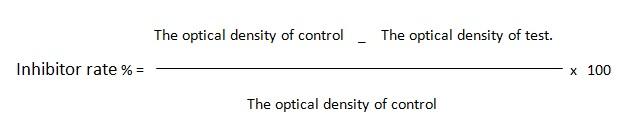
Cytotoxicity of Blessed Thistle Blooms Extraction: The study results on the cytotoxicity of a plant extract on cervical cancer cells exhibited a considerable elevation in the inhibition of cancer cell growth as the concentration of the extract and the duration of incubation increased. In other words, the growth inhibition obeyed a pattern that was dependent on both the concentration and time (Table 1 and Figure 1).
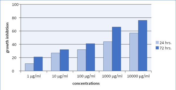
Figure 1: Effect Different Concentrations of Plant Extracts and Incubation Period on the Proliferation of Cervical Cancer Cells
Table 1: Effect of Different Concentrations of Plant Extracts and Incubation Period on the Proliferation of Cervical Cancer Cells
|
Concentration µg/mL |
24 |
72 |
p-value |
|
1 |
11 E |
21 C |
0.0001* |
|
10 |
27 D |
32 BC |
0.04* |
|
100 |
32 C |
41 B |
0.0001* |
|
1000 |
44 B |
66 A |
0.0001* |
|
10000 |
57 A |
76 A |
0.0001* |
|
LSD |
3.64 |
15.44 |
- |
|
IC 50 µg/ml |
8196.7 µg/ml |
6097.6 µg/ml |
- |
unlike capital letters exhibit significant differences (p<0.05) among means of the respective columns, * Significant at (p≤0.05)
Cytotoxicity of Vinblastine
The study results on the cytotoxic marks of vinblastine on cervical cancer cells exhibited a significant rise in the suppression of cancer cell multiplication with longer incubation periods and higher concentrations. The growth inhibition especially exhibited a temporal dependency (Table 2 and Figure 2).
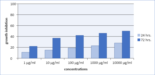
Figure 2: Impact of Diverse Concentrations and Incubation Durations of Vinblastine on the Multiplication of Cervical Cancer Cells
Table 2: Impact of Diverse Concentrations and Incubation Durations of Vinblastine on the Multiplication of Cervical Cancer Cells
|
Concentration µg/mL |
24 |
72 |
p-value |
|
1 |
11D |
22 C |
p<0.001* |
|
10 |
15 CD |
37 B |
p<0.001* |
|
100 |
19 BC |
42 AB |
p<0.001* |
|
1000 |
23 AB |
46 AB |
p<0.001* |
|
10000 |
28 A |
50 A |
p<0.02* |
|
LSD |
7.1 |
9.2 |
- |
|
IC 50 µg/mL |
16666.7 |
9091 |
- |
unlike capital letters exhibit significant differences (p<0.05) among means of the respective columns, * Significant at (p≤0.05)
The Cytotoxicity of a Combination of Plant Extract and Vinblastine
Study outcomes display the capability of the combination to attenuate cancer cell growth; the pattern of growth inhibition shows a significant elevation in the growth inhibition of cancer cells with an elevation in the combination concentration and period of incubation (time and concentration dependent) (Table 3 and Figure 3).
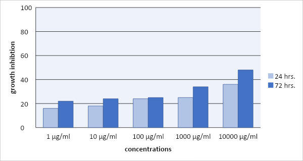
Figure 3: Outcomes of Diverse Concentrations and Durations of Incubation of a Mixture of Vinblastine and Plant Extract on the Multiplication of Hela Cell Line
Table 3: Outcomes of Diverse Concentrations and Durations of Incubation of a Mixture of Vinblastine and Plant Extract on the Multiplication of Cervical Cancer Cells
|
Concentration µg/ml |
24 |
72 |
p-value |
|
1 |
16 B |
22 C |
p<0.010* |
|
10 |
18 B |
24 BC |
p<0.065 |
|
100 |
24 B |
25 BC |
p<0.800 |
|
1000 |
25 AB |
34 B |
p<0.072 |
|
10000 |
36 A |
48 A |
p<0.001* |
|
LSD |
11.16 |
12.82 |
- |
|
IC 50 µg/ml |
13157.9 |
9801 |
- |
unlike capital letters exhibit significant differences (p<0.05) among means of the respective columns, * Significant at (p≤0.05)
Drug Combination Studies
The combination study between vinblastine and plant extract indicated the subsequent outcomes. At 24 incubation periods, a concentration of 1 µg/ml for the combination exhibited potent synergism, while other concentrations showed antagonistic impact. At 72 incubation periods, there was an antagonism impact between the combination components for all concentrations.
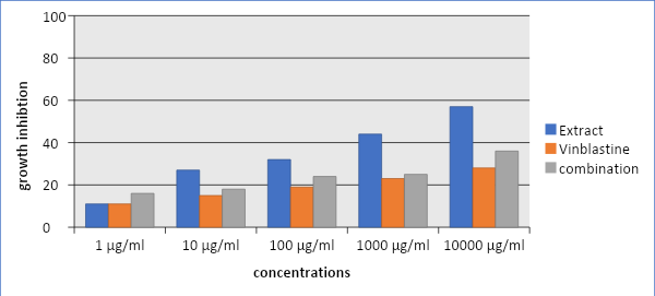
Figure 4: Compare the Influence of Extract, Vinblastine and a Mixture on the Cytotoxicity of the Hela Cell Line During a 24-hour Incubation Period
Table 4: Compare the Influence of Extract, Vinblastine and a Mixture on the Cytotoxicity of the Hela Cell Line During a 24-hour Incubation Period
|
Comparison 24 hrs. |
||||
|
Concentration µg/ml |
Extract |
Vinblastine |
combination |
LSD |
|
1 |
b 11 E |
b 11 D |
a 16 B |
4 |
|
10 |
a 27 D |
b 15 CD |
b 18 B |
5.66 |
|
100 |
a 32 C |
b 19 BC |
ab 24 B |
9.78 |
|
1000 |
a 44 B |
b 23 AB |
b 25 AB |
13.64 |
|
10000 |
a 57 A |
c 28 A |
b 36 A |
6.92 |
|
LSD |
3.64 |
7.1 |
11.16 |
|
|
IC 50 µg/ml |
8196.7 |
16666.7 |
13157.9 |
|
Different capital letters exhibit substantial disparities (p<0.05) among the averages of the corresponding columns. Different lowercase letters mark significant variances (p<0.05) among the averages of the corresponding rows
Based on the dose reduction index, the cytotoxicity of the combination at a concentration of 1 μg/mL was estimated after 24 hours of incubation, revealing a significant reduction in the needed dosage for both vinblastine and the extract at this concentration, displaying a favorable dose reduction. Regardless, other concentrations did not reveal a favorable dose reduction. At a 72-hour incubation time, the dose reduction index value for the mixture exhibits an unfavorable dosage reduction between the combination elements in all concentrations (Table 6,7).
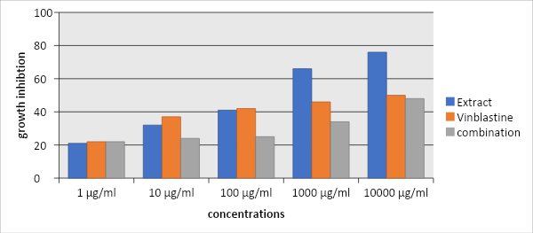
Figure 5: Compare the Influence of Extract, Vinblastine and a Mixture of Cytotoxicity on the Hela Cancer Cell Line During a 72-hour Incubation Period
Table 5: Compare the Influence of Extract, Vinblastine and a Mixture on the Cytotoxicity of the Hela Cell Line During a 72-hour Incubation Period
|
Comparison 72 hrs. |
||||
|
Concentration µg/ml |
Extract |
Vinblastine |
combination |
LSD |
|
1 |
a 21 C |
a 22 C |
a 22 C |
N. S |
|
10 |
ab 32 BC |
a 37 B |
b 24 BC |
9.78 |
|
100 |
a 41 B |
a 42 AB |
b 25 BC |
12.64 |
|
1000 |
a 66 A |
b 46 AB |
c 34 B |
9.78 |
|
10000 |
a 76 A |
b 50 A |
b 48 A |
11.98 |
|
LSD |
3.64 |
9.2 |
12.82 |
- |
|
IC 50 µg/ml |
6097.6 |
9091 |
9801 |
- |
Different capital letters exhibit substantial disparities (p<0.05) among the averages of the corresponding columns. Different lowercase letters mark significant variances (p<0.05) among the averages of the corresponding rows
DISCUSSION
A separate study confirmed our research findings, providing evidence that the alcoholic extract obtained from the blossoms of Cnicus Benedictus might inhibit the proliferation of breast cancer cells in a controlled laboratory environment [10]. Others exhibit that the ethyl extract of blessed thistle can inhibit the growth of Dalton's lymphoma ascites cells [19].
The main determinant of the anticancer effects of plant extract is its phytochemical contents, particularly flavonoids. Flavonoids have been discovered to have an inhibitory impact on the growth of many kinds of cancer cells, comprising the constituents found in the blood, brain, lung, uterus, salivary gland and melanoma. This phenomenon is more evident in cells with a high mitotic index as opposed to those with a low mitotic index [20].
Table 6: The Combined Effect of Vinblastine and a Plant Extract on Hela Cancer Cells After 24-Hour Incubation Period
|
Dose reduction index value |
Pattern of combination |
CI value |
Con. ratio |
Drug μg/ml |
||
|
extract |
vinblastine |
1:1 |
extract |
vinblastine |
||
|
4.64430 |
48.8329 |
Strong Synergism |
0.23580 |
0.5 μg/ml |
0.5 μg/ml |
|
|
0.84130 |
15.5611 |
Moderate Antagonism |
1.25291 |
5 μg/ml |
5 μg/ml |
|
|
0.38577 |
30.3508 |
Antagonism |
2.62518 |
50 μg/ml |
50 μg/ml |
|
|
0.04838 |
4.72037 |
Very Strong Antagonism |
20.8821 |
500 μg/ml |
500 μg/ml |
|
|
0.04328 |
33.9019 |
Very Strong Antagonism |
23.1369 |
5000 μg/ml |
5000 μg/ml |
|
The CI and DRI values were analyzed using Compusyn software, The CI and DRI values were analyzed using Compusyn software; A CI value more than 1 denotes antagonism, a CI equal to 1 exhibit an additive effect and a CI less than 1 indicates synergism. A dose reduction index (DRI) more than one is correlated with less toxicity
However, phenolic chemicals can inhibit the growth of breast cancer cells. activation of the apoptotic pathway was the mechanism that inhibited cell proliferation in MDA-MB-231 [21].
The anticancer activities of Cnicus Benedictus L. flower are thought to stem from its osmolality impact. Extracts have a rich supply of proteins, carbohydrates, minerals and other substances that cause the environment of cancer cells to become hypertonic. This leads to an inhospitable environment for the cancer cells and may potentially cause a dose-dependent osmotic shock to the cell lines [22].
furthermore, our study outcomes on the cytotoxicity of vinblastine align with the outcomes of another investigation as presented, Since the cytotoxic effects of vinblastine seem to be affected by both the quantity of the medicine and the period of exposure, it is evident that female uterine cervical cancer cell lines are susceptible to different concentrations of vinblastine. The mechanism of action of vinblastine is governed by its cytotoxic effects, which become apparent after a 72-hour incubation period. The observed effects are explained by the prolonged presence of vinblastine inside cancer cells, leading to a significant increase in cell death. 28 different lymphoid cancers may be induced to generate microtubule crystals by Vinblastine. The length of vinblastine therapy has a significant impact on both the number of cells exhibiting microtubule abnormalities and the extent of their progression [23-25].
Table 7: The Combined Effect of Vinblastine and a Plant Extract on Hela Cancer Cells After 72-Hour Incubation Period
|
Dose reduction index value |
Pattern of combination |
CI value |
Con. ratio |
Drug μg/ml |
||
|
extract |
vinblastine |
1:1 |
extract |
vinblastine |
||
|
3.24724 |
0.36375 |
Antagonism |
3.05706 |
0.5 μg/ml |
0.5 μg/ml |
|
|
0.48846 |
0.08701 |
Very Strong Antagonism |
13.5407 |
5 μg/ml |
5 μg/ml |
|
|
0.05938 |
0.01321 |
Very Strong Antagonism |
92.5596 |
50 μg/ml |
50 μg/ml |
|
|
0.02863 |
0.03802 |
Very Strong Antagonism |
61.2308 |
500 μg/ml |
500 μg/ml |
|
|
0.02356 |
0.34286 |
Very Strong Antagonism |
45.3677 |
5000 μg/ml |
5000 μg/ml |
|
The CI and DRI values were analyzed using Compusyn software; A CI value more than 1 denotes antagonism, a CI equal to 1 exhibits an additive effect and a CI less than 1 indicates synergism. A dose reduction index (DRI) more than one is correlated with less toxicity
Based on the study's results, it can be assumed that when the combination is employed at the greatest concentration, it leads to a declined degree of growth inhibition corresponding to the cytotoxicity of the plant extract sole after 24 hours. After 72 hours, the combination of plant extracts and vinblastine resulted in a lessened level of growth inhibition compared to the individual plant extracts and vinblastine alone.
According to the combination index data, combination displays antagonistic manner at all concentrations except the lowest one, which may be attributed to the ability of vinblastine to influence the fluidity of cellular membranes [26]. This may result in the retardation of the transportation of the plant extract components over the cellular membrane into the cell, this phenomenon mainly displays at high concentrations of vinblastine.
The synergistic pattern becomes observable at a concentration of 1µg/ml. At smaller concentrations, vinblastine has a diminished influence on membrane fluidity, resulting in a reduced ability to prevent the access of plant extract components into the cell [27,28].
Vinblastine targets spindle microtubules and the plant extract displays a comparable target by selectively targeting cells with a high mitotic index rather than those with a lower mitotic index [20]. This mechanism possibly clarifies the synergistic manners seen between vinblastine and the elements of the plant extract.
A limitation of our study was the exclusion of cytotoxicity data within a 48-hour timeframe, this period does not supply us with a reliable indication of the pattern of cytotoxicity, whether it is concentration-dependent or time-dependent.
CONCLUSIONS
The study results revealed to, the Cnicus Benedictus L. flower extract exhibited capability to attenuate cervical cancer cell growth by (cycle and non-Cyle specific) manner of cytotoxicity. In contrast, the mixture of vinblastine and Cnicus Benedictus L. flower extract showed two distinct stages of cytotoxicity on cervical cancer cells. Synergism was mostly seen at lower doses during the first phase, while the second phase exhibited mostly antagonism at moderate and higher doses.
Author Contributions
- Creation and Development: Marwan I. Al-Zuhairi, Diyar M. jalil
- Data Collection and Organization: Sadeq Jaafer Al-Tameemi
- Data Analysis and Interpretation: Marwan I. Al-Zuhairi
- Composition of the Article: Diyar M. jalil
- Critical Revision of Article for Important Intellectual Content: Marwan I. Al-Zuhairi
- Proficiency in Statistical Analysis: Diyar M. jalil, Sadeq Jaafer Al-Tameemi
- Ultimate Endorsement and Guarantee of the Article: Sadeq Jaafer Al-Tameemi, Marwan I. Al-Zuhairi
Acknowledgement
The faculty and staff of al-Mustansiriyah University and the ICMGR, Iraq's Centre for Cancer and Medical Genetics in Bagdad, Iraq, played a crucial role in assisting me with my research. I want to express my sincere gratitude to all of them.
REFERENCES
- Wang, S. et al. “Travelers’ food experience sharing on social network sites.” Journal of Travel & Tourism Marketing, vol. 34, no. 5, 2017, pp. 680–693.
- Deo, S.V.S. et al. “GLOBOCAN 2020 report on global cancer burden: Challenges and opportunities for surgical oncologists.” Annals of Surgical Oncology, vol. 29, no. 11, 2022, pp. 6497–6500.
- Omone, O.M. et al. “Effective prevention of high-risk cervical cancer among women in developing countries: a case study in Nigeria.” 2020.
- Rock, C.L. et al. “American cancer society guideline for diet and physical activity for cancer prevention.” CA: A Cancer Journal for Clinicians, vol. 70, no. 4, 2020, pp. 245–271.
- Mustapha, A. et al. “Cancer chemotherapy: A review update of the mechanisms of actions, prospects and associated problems.” J Biomed, vol. 1, no. 1, 2022, pp. 1–16.
- Shoji, T. et al. “Hepatitis B virus reactivation during temozolomide administration for malignant glioma.” International Journal of Clinical Oncology, vol. 26, 2021, pp. 305–315.
- Batziakas, K.G. et al. “Reducing preharvest food losses in spinach with the implementation of high tunnels.” Scientia Horticulturae, vol. 265, 2020, p. 109268.
- Wied, J.P. et al. “Invasive grasses in south texas rangelands: historical perspectives and future directions.” Invasive Plant Science and Management, vol. 13, no. 2, 2020, pp. 41–58.
- Jian, Y.L. et al. “Flavonoid compositions and antioxidant activity of immature kumquat in different flowering periods.” Taiwanese Journal of Agricultural Chemistry & Food Science, vol. 58, 2020.
- Al-Samarray, Y.S. et al. “The cytotoxic effect of ethanolic extract of cnicus benedictus L. flowers on the murine mammary adenocarcinoma cancer cell line AMN-3.” Biochemical & Cellular Archives, vol. 20, 2020.
- Yasin, Y.S. et al. “Effect of laetrile vinblastine combination on the proliferation of the hela cancer cell line.” Asian Pacific Journal of Cancer Prevention, vol. 24, no. 12, 2023, pp. 4329–4337.
- Jumaa, A.H. “The Cytotoxic effect of vincristine-amygdalin combination on human cervical cancer cell line (HeLa Cancer Cell Line).” Iraqi Journal of Cancer and Medical Genetics, vol. 9, no. 2, 2016.
- Jumaa, A.H. et al. “Esomeprazole and amygdalin combination cytotoxic effect on human cervical cancer cell line (HeLa Cancer Cell Line).” Journal of Pharmaceutical Sciences and Research, vol. 10, no. 9, 2018, pp. 2236–2241.
- De Sousa, K.P. et al. “Isolation and characterization of extracellular vesicles and future directions in diagnosis and therapy.” Wiley Interdiscip Rev Nanomed Nanobiotechnol, vol. 15, 2023, pp. e1835.
- Ueda, K. et al. “Analysis of the complete genome sequences of clostridium perfringens strains harbouring the binary enterotoxin BEC gene and comparative genomics of pCP13-like family plasmids.” BMC Genomics, vol. 23, 2022, pp. 226.
- Ayiomamitis, G.D. et al. “Understanding the interplay between COX-2 and HTERT in colorectal cancer using a multi-omics analysis.” Cancers (Basel), vol. 11, 2019, pp. 1536.
- Meyer, C.T. et al. “Charting the Fragmented Landscape of Drug Synergy.” Trends in Pharmacological Sciences, vol. 41, no. 4, 2020, pp. 266–280.
- Bailer, A.J. Statistical Programming in SAS. CRC Press, 2020.
- Human, B. MDA-MB-468/MCF7/MDA MB-231/SK-BR-3 C-33 A. Alternative Remedies and Natural Products for Cancer Therapy: An Integrative Approach, 2023, pp. 82.
- Khan, H. et al. “Flavonoids nanoparticles in cancer: Treatment, prevention and clinical prospects.” Seminars in Cancer Biology, vol. 69, February 2021, pp. 200–211.
- Roleira, F.M. et al. “Phenolic derivatives from medicinal herbs and plant extracts: Anticancer effects and synthetic approaches to modulate biological activity.” Studies in Natural Products Chemistry, vol. 57, 2018, pp. 115–156.
- Ijarotimi, O.S. et al. “Nutritional, antioxidant, angiotensin-converting-enzyme and carbohydrate-hydrolyzing-enzyme inhibitory activities of underutilized leafy vegetables: african wild lettuce (Lactuca Taraxacifolia Willd).” Clinical Phytoscience, vol. 7, no. 1, 2021, pp. 1–13.
- Kadhim, R.S. and S.A. Mohamad. “Comparison of laparoscopic and conventional surgery of unilateral ovariectomy in jennies.” Acta Biomed, vol. 94, no. 2, 2023, pp. e2023140.
- Della Corte, L. et al. “Advances in paclitaxel combinations for treating cervical cancer.” Expert Opinion on Pharmacotherapy, vol. 21, no. 6, 2020, pp. 663–677.
- Škubník, J. et al. “Vincristine in combination therapy of cancer: Emerging trends in clinics.” Biology, vol. 10, no. 9, 2021, pp. 849.
- Soteriou, C. et al. “Advances in understanding and in multi-disciplinary methodology used to assess lipid regulation of signalling cascades from the cancer cell plasma membrane.” Progress in Lipid Research, vol. 81, 2021, pp. 101080.
- Tcheng, M. et al. “Structure defines bioactivity of avocado-derived acetogenins.” Studies in Natural Products Chemistry, vol. 78, 2023, pp. 1–44.
- Negi, A.S. and S. Jain. “Recent advances in natural product-based anticancer agents.” Studies in Natural Products Chemistry, vol. 75, 2022, pp. 367–447.