
Journal of Advanced Biological Sciences
ISSN Print: N/A
ISSN Online: 3134-8823
About: Journal of Advanced Biological Sciences (JABS) is a peer-reviewed, open-access journal that aims to publish cutting-edge research and advancements in all areas of biological sciences. The journal serves as a platform for researchers, academicians, and professionals to contribute their scientific knowledge and foster innovation in biology-related fields.
Journal of Advanced Biological Sciences | Year 2024 | Volume 1 | Issue 1 | Pages 22-33
Endophytes and the Evolution of Identification Methods: A Comprehensive Review
Fahmida Khatoon 1*1Department of Biochemistry, College OF Medicine, University of Ha’il, Saudi Arabia
2Department of Nursing, College of Nursing and Health Sciences, Jazan University, Saudi Arabia
View PDF Download XML Download DOI XML DOI: 10.66590/jabs2024010104
Abstract
Endophytes are a unique group of microorganisms that reside within plant tissues without causing any apparent harm to their host. The present review focuses on various aspects of endophytic research conducted in the Kingdom of Saudi Arabia over the past decade. It highlights the diversity of endophytes isolated from native and medicinal plants adapted to arid and semi-arid environments. Furthermore, this review provides a comparative analysis of different methodologies used for the identification of endophytes, emphasizing the transition from traditional techniques to modern molecular approaches. Advanced tools such as DNA barcoding, Random Amplified Polymorphic DNA (RAPD) analysis and sequence-based identification using BLAST have been increasingly applied in recent years for accurate characterization of endophytes. A compilation of commonly reported endophytic species isolated from various plant hosts across different regions of Saudi Arabia is also presented. The rich floral diversity of the region, including desert-adapted and medicinal plants, represents a valuable source of bioactive compounds as well as endophytic microorganisms with significant biotechnological potential. Given the unique ecological conditions of Saudi Arabia, further exploration and identification of endophytes from indigenous plant species are of great importance. Such studies may contribute to advancements in pharmaceuticals, agriculture and environmental sustainability, ultimately benefiting human health and scientific development.
INTRODUCTION
The living world is characterized by immense biological diversity, which plays a fundamental role in sustaining ecosystems and maintaining ecological balance. Interactions between microorganisms, plants and animals are critical components of this balance. Among these interactions, endophytism where microorganisms reside within plant tissues without causing harm has gained significant attention due to its wide-ranging ecological and biotechnological importance.
Endophytes have emerged as a promising resource in various sectors, including agriculture, pharmaceuticals and biotechnology. Their potential applications range from the production of biofuels to the synthesis of pharmacologically active compounds, making them valuable for industrial and medical research. Previous studies have demonstrated that endophytes can serve as a source of novel bioactive metabolites with antimicrobial, anticancer and anti-inflammatory properties.
Despite the growing interest, the exploration and identification of endophytes remain challenging due to their immense diversity and often cryptic nature within host plants. Many endophytes remain undetected because of limitations in conventional identification techniques. Furthermore, the diversity, abundance and distribution of endophytic communities are highly dynamic and influenced by multiple abiotic factors such as climate, soil conditions and water availability, as well as biotic factors including host plant species and microbial interactions. Endophytes are distinct from epiphytes in that they inhabit internal plant tissues, whereas epiphytes exist on the external surfaces of plants and may sometimes exhibit pathogenic behavior. In contrast, endophytes typically maintain a symbiotic or neutral relationship with their host.
This review focuses on the diversity and identification of endophytes in the Kingdom of Saudi Arabia over the past decade, with particular emphasis on native and medicinal plants adapted to arid and semi-arid environments. Saudi Arabia’s unique ecological conditions and rich plant diversity provide an excellent opportunity for discovering novel endophytic microorganisms with significant biotechnological potential. However, research on endophytes associated with indigenous medicinal plants in this region remains limited.
Therefore, there is a pressing need for comprehensive studies to explore, isolate and identify endophytes from Saudi Arabia’s native flora. Such efforts could contribute significantly to advancements in pharmaceuticals, agriculture and environmental sustainability, ultimately benefiting human health and scientific progress.
Origin of the Term ‘Endophyte’
The term endophyte is derived from the Greek words endon (within) and phyton (plant), referring to organisms that live inside plant tissues and complete part or all of their life cycle within the host. Endophytes were first described by the German botanist Johann Heinrich Friedrich Link in 1809. Initially, these organisms were considered parasitic fungi and were later referred to as “microzymas.” In 1887, Victor Galippe reported the presence of bacteria within plant tissues, expanding the concept of endophytes to include diverse microbial groups.
Over time, several definitions of endophytes have been proposed. Petrini described endophytes as organisms that colonize internal plant tissues without causing harm. Similarly, Stone et al. [1], defined them as microorganisms that inhabit plant tissues asymptomatically for at least a part of their life cycle. Jonathan R. Gaiero et al. further characterized endophytes as bacterial or fungal species that can be isolated from surface-sterilized plant tissues and do not negatively affect plant growth.
Endophytes exhibit remarkable diversity and may include bacteria, fungi, algae and actinomycetes. These organisms reside within plant tissues without inducing visible disease symptoms under normal conditions. Some actinomycetes have been identified as important endophytes in medicinal plants due to their ability to produce bioactive compounds. Additionally, endophytes have been reported in association with lichens, highlighting their adaptability across different ecological niches.
Under certain environmental conditions, some endophytes may behave as latent pathogens, becoming pathogenic when the host plant experiences stress. Such stress factors may include abiotic conditions such as drought and temperature extremes, as well as biotic pressures like inter-plant competition. This is particularly relevant in arid and semi-arid regions such as the Kingdom of Saudi Arabia, where environmental stressors are prominent.
Endophytes are also considered niche-specific organisms, with their diversity influenced by host plant species and habitat type. They have been isolated from a wide range of plant hosts, including medicinal plants, hydrophytes, mesophytes and xerophytic species. In extreme environments, such as desert ecosystems, endophytic fungi have demonstrated thermotolerance and the ability to confer stress resistance to their host plants. This mutualistic relationship is especially significant in desert flora, where endophytes may enhance plant survival under harsh climatic conditions.
Given the unique environmental conditions and rich plant diversity of Saudi Arabia, the study of endophytes in this region offers significant potential for discovering novel microorganisms with important ecological and biotechnological applications.
Life Cycle of Endophytes
The life cycle of endophytes typically begins with the initial infection of the host plant, often at the seed stage. As the seed germinates and develops into a mature plant, the endophytes colonize internal tissues and establish a stable association with the host. During plant growth, these microorganisms proliferate and spread to various plant parts, including roots, stems and leaves.
With the progression to the reproductive stage, endophytes may be transmitted to new host plants through vertical transmission via seeds or horizontal transmission through pollen. This ensures the persistence and dissemination of endophytic populations across plant generations. The life cycle of endophytes is commonly illustrated through diagrammatic representations (Figures 1 and 2).
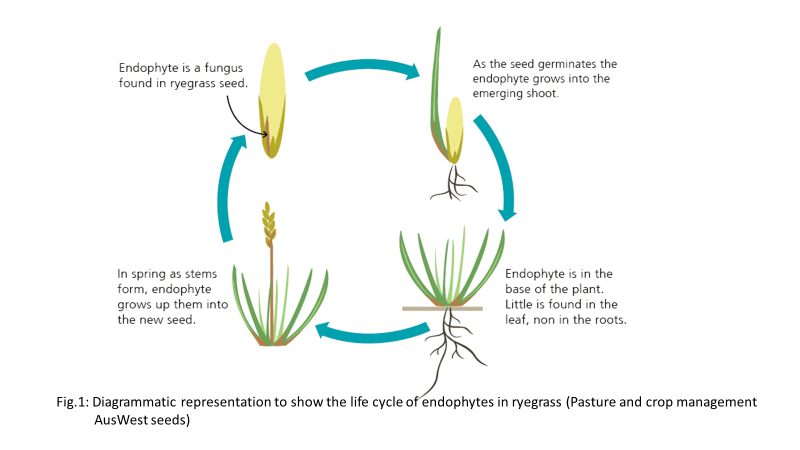
Figure 1: Diagrammatic Representation to Show the Life Cycle of Endophytes in Ryegrass (Pasture and Crop Management AusWest Seed)
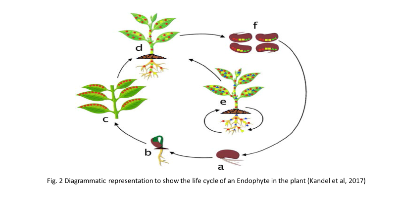
Figure 2: Diagrammatic Representation to Show the Life Cycle of Endophyte in the Plant [3]
Types and Diversity of Endophytes
Endophytes can be broadly classified based on the type of microorganisms inhabiting host plants into bacterial, fungal, cyanobacterial and actinomycete endophytes. Among these, bacterial and fungal endophytes are the most extensively studied groups.
Bacterial Endophytes
Bacterial endophytes are widely distributed and have been reported to colonize almost all plant tissues, including roots, stems, leaves and reproductive structures. Their colonization is influenced by the mutual benefits derived from the host plant. The primary entry points for these microorganisms are natural openings and wounds formed during plant growth and development.
Common genera of bacterial endophytes include Azospirillum, Bacillus, Methylobacterium and Pseudomonas. These bacteria play a crucial role in plant growth promotion by enhancing nutrient uptake, producing phytohormones and improving stress tolerance. Additionally, they act as biological control agents by protecting host plants against pathogens and herbivores. Both Gram-positive, Gram-negative bacteria and actinobacteria have been reported as endophytes.
Fungal Endophytes
Fungal endophytes are highly diverse and ubiquitous in nature. Common genera include Alternaria, Fusarium and Colletotrichum. These fungi often establish mutualistic relationships with host plants, enhancing resistance to herbivores, pathogens and environmental stress. In many cases, endophytic fungi enable plants to produce secondary metabolites with antimicrobial or toxic properties, which contribute to plant defense mechanisms.
Actinomycetes and Other Endophytes
Endophytic actinomycetes have also gained attention due to their ability to produce bioactive compounds with antimicrobial and pharmaceutical importance. Many of these organisms exhibit strong antagonistic activity against plant pathogens. In addition, endophytes have been reported in association with macroalgae and lichens, highlighting their ecological adaptability across diverse environments.
Factors Influencing Endophytic Diversity
The type and diversity of endophytes present within a host plant are influenced by multiple factors, including plant species, developmental stage, physiological condition and environmental parameters such as soil type, temperature, altitude and water availability. In arid and semi-arid regions like Saudi Arabia, these factors play a particularly significant role in shaping endophytic communities.
Endophytes typically engage in symbiotic relationships with their host plants, where the plant provides nutrients and protection, while the microorganisms enhance plant growth and stress tolerance. However, some endophytes may exhibit “balanced antagonism,” shifting between mutualistic and parasitic behavior depending on environmental conditions.
Endophytic communities may be either host-specific or ubiquitous. Environmental conditions surrounding the host plant significantly influence fungal colonization patterns. Moreover, plants especially medicinal species are rich in secondary metabolites such as flavonoids, phenols, saponins and essential oils, which create a favorable environment for endophyte survival and proliferation.
Relationship of Endophytes with the Host Plant
Endophytes shows life style variations as they have been found in different forms in the plant [2]. They may be found as symbionts, saprotrophs, latent pathogens and as Diazotrophic endophytes (Where nitrogen fixation occurs without root nodules) Doty S et al. ]3]. According to Ahmad R et al [4], latent pathogens known to live without showing symptoms in the host plant having an epiphytic phase in their life cycle are also endophytes. Endophytes can invade any part of the plant and can grow there. The relationship that they establish varies from symbiotic to pathogenic. Much has been studied about the interactions between the endophytes and the host plant. In certain cases, the interactions can involve more than two partners. Endophytic bacteria and fungi may interact not only with the plant host, but also with other organisms, including mycorrhizal fungi and metazoa [5]. They also explained the life history variations shown by the endophytes. It refers to the different stages or forms in which the endophytes can interact with the host plant.
On the basis of the association of the endophytes with the host plant following three types of categories can be made:
- Endophytes as Commensals: Fungal endophytes of plants are known to behave as commensals. Commensalism is an interaction where one of the species is benefitted and the another species is neither benefitted nor harmed. The organisms showing this kind of interaction are called as commensals. In this kind of interaction, the endophytes, both fungi as well as the bacteria harbour the different parts of the plants to derive nutrition from them, but do not benefit the host plant in any way. Commensalism of endophytes has been studied and established by Creman R et al. in the endophytes of locoweeds
- Endophytes as Symbionts: Endophytes have been found to be having symbiotic or mutualistic relationship with the host plant. In this kind of association both the endophyte and the host plant are at benefit. For example, it has been found that in many cases because of the type of the endophytic species found in the host plant, different kind of secondary metabolites are produced, giving defence mechanism to the host plant. Thus the host plants are protected against herbivory. In return the endophytes derive nutrition from the host plant and occupy space within it. For example, AMF (Arbuscular Mycorrhizal Fungi) is one such endophyte found in the plant Glomeromycota. Mutualistic interaction of both fungi and bacteria as endophytes benefit them by supplying nutrients from the host plant as well as by providing protection from the environmental stresses [5]
- Endophytes as Parasites: Very little is known regarding the pathogenic effect of endophytes on the host plant. Under normal growth conditions endophytes can have detrimental effect on the host plant. One such example is Fusarium verticilloides. One of the reason stated for endophytes to behave as parasites is the abiotic stress and reduced fitness of the host plant. In such conditions the endophytes may produce such chemicals or toxins which can be harmful for the host plant. Studies have shown that endophytes can behave as parasites only when the balance between the two partners that is the host and the endophytic microbe somehow gets disturbed. It may be due to environmental stress or physiological or due to some genetic reasons [6]. Bernard S et al [7], have also mentioned the endophytes to behave as latent pathogens under stressful environmental conditions. However how this stress full environmental condition can bring about changes in the behaviour of the endophytes towards the host plant needs further study. It has also been stated that some fungal endophytes can shift between parasitic and mutualistic life strategies, known as balanced antagonism
According to Rodriguez et al [8], Endophytic fungi are classified into two major groups- Clavicipitaceous endophytes which infect some grasses. Non clavicipitaceous endophytes which are found within other plant groups. Clay and Schardl recognized three types of Clavicipitaceous endophytes (C-endophytes)-Type I which include symptomatic and pathogenic species. Type II and Type III which include mixed interaction and asymptomatic endophytes. Fungal endophytes can again be categorised into four major or broad groups based on host range, tissue colonized, their mode of transmission etc [8]. Class I include Clavicipitaceous endophytes whereas Non-clavicipitaceous endophytes include class II, III and IV. Class I endophytes basically belong to class Ascomycota and are limited to some cool and warm season grasses. They are found within plant shoots, where they form intercellular infections. They have been reported to increase plant biomass, provide drought tolerance and produce chemicals that are toxic to animals and thus decrease herbivory. Class II endophytes include a diversity of species, all of which are members of Dikarya (Ascomycota or Basidiomycota). They have been reported to confer habitat specific stress tolerance to host plant. Class III endophytes are highly diverse within individual host tissues, plants and populations. They are found to be different from other class of endophytes on the basis of their occurrence in above- ground tissues, horizontal transmission, formation of highly localized infections. Class IV endophytes are the ones having darkly melanized septa and are restricted to plant roots. They are prevalent in high stress environment.
Other than Bacteria and Fungi, Cyanobacteria have also been found as endophytes of several plant species, specially the lower forms of plants like moss, liverworts and ferns. In some species of Orchids [9], they have been found to be in intimate relationship. In their relationship, the cyanobacteria species help the plant in acquiring the fixed nitrogen from the atmosphere. In return Cyanobacteria gets a stable environment and the wider nutrient source available from the host. Although their occurrence in the plants are less studied or isolated but their absence in the host plants cannot be accepted.
Actinomycetes have also been isolated from medicinal plants growing in different parts of India [10,11]. They have been mostly isolated from root tissue followed by stem and then rhizome.
Entry and Colonization of Endophtes in The Host
Endophytes exhibit diverse lifestyles and can exist in multiple functional forms within plant tissues. They may occur as symbionts, saprotrophs, latent pathogens, or diazotrophic endophytes capable of nitrogen fixation without the formation of root nodules. Their ability to colonize various plant parts including roots, stems, leaves and reproductive structures highlights their ecological versatility. The nature of their interaction with the host plant can range from mutualistic to pathogenic, depending on environmental and physiological conditions.
Endophytes may also participate in complex, multi-partner interactions involving not only the host plant but also other microorganisms such as mycorrhizal fungi and even higher organisms. These interactions reflect the dynamic life history strategies of endophytes, which may shift across different stages of their lifecycle.
Types of Endophytic Associations
Based on their interaction with the host plant, endophytes can be categorized into three main groups:
- Commensal Endophytes: In commensal relationships, endophytes derive nutrients and shelter from the host plant without causing harm or providing significant benefits. Both bacterial and fungal endophytes may exist in this form, colonizing plant tissues passively
- Symbiotic (Mutualistic) Endophytes: In mutualistic associations, both the endophyte and the host plant benefit. Endophytes may enhance plant growth, improve nutrient acquisition and confer resistance to environmental stress and herbivory. In return, the plant provides nutrients and a protective niche. For example, Arbuscular Mycorrhizal Fungi (AMF) are well-known mutualistic endophytes that enhance plant nutrient uptake and stress tolerance
- Parasitic Endophytes: Under certain conditions, endophytes may exhibit pathogenic behavior. Environmental stress, reduced host fitness, or disruption in host–microbe balance can trigger this shift. In such cases, endophytes may produce toxic metabolites that harm the plant. This phenomenon, known as “balanced antagonism,” reflects the ability of some endophytes to switch between mutualistic and parasitic lifestyles
Fungal Endophyte Classification
Fungal endophytes are broadly classified into two major groups:
Clavicipitaceous Endophytes (C-endophytes)
These primarily infect grasses and are often associated with systemic colonization. They are further subdivided into:
- Type I: Pathogenic and symptomatic
- Type II & III: Mixed or asymptomatic interactions
Non-Clavicipitaceous Endophytes
These are more diverse and occur in a wide range of plant species. They are further divided into four classes based on host range, colonization pattern and transmission mode:
- Class I: Limited to grasses, systemic infection, often beneficial (e.g., drought tolerance, herbivore resistance)
- Class II: Broad host range, provide habitat-specific stress tolerance
- Class III: Highly diverse, localized infections in above-ground tissues, horizontally transmitted
- Class IV: Root-associated, darkly pigmented fungi, common in high-stress environments
Other Types of Endophytes
In addition to bacteria and fungi, other microorganisms such as cyanobacteria and actinomycetes also function as endophytes. Cyanobacterial endophytes are commonly associated with lower plants like mosses, liverworts and ferns and play a significant role in nitrogen fixation. In certain plant species, including orchids, they form intimate symbiotic relationships, providing fixed nitrogen in exchange for nutrients and shelter.
Endophytic actinomycetes have also been widely studied, particularly in medicinal plants. These organisms are predominantly isolated from root tissues and are known for producing bioactive compounds with antimicrobial properties.
Ecological Significance
Endophytic communities within a host plant may be either host-specific or widely distributed across different plant species. Their composition is strongly influenced by environmental conditions, especially in extreme ecosystems such as arid and semi-arid regions. These microorganisms contribute significantly to plant adaptation, survival and productivity under stress conditions.
The preferred site of entry for the bacterial endophytes are the regions having thin surface such as root hairs or the elongation zone of the apical root meristem. In leaf colonization, bacterial strains are firstly attached to the surface of leaf and get randomly distributed throughout. They may enter the leaf tissue via natural openings such as stomata, hydathodes and influence their local environment [12]. It has been observed that the bacterial endophytes use their cellular modifications like flagella, fimbriae or cell surface polysaccharides for attachment to the plant surface. Bacterial endophytes occupy intercellular spaces in the plant as these areas have abundance of carbohydrates, amino acids and inorganic nutrients. In leaves bacterial endophytes have been observed in the intercellular spaces of mesophyll, xylem tissue and substomatal areas [3]. As far as colonization in seed is concerned, it occurs in the different parts of the seed including the embryo (Figure 3).
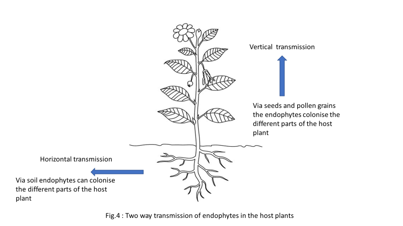
Figure 3: Two Way Transmission of Endophytes in the Host Plants
Variation in Colonization
Endophytes colonize in all most all the parts of the plant as shown in Figure 4. Colonization frequency and isolation frequency has been found to be greater in leaves than the bark tissue [13]. But dense colonization of leaves by endophytes affect photosynthesis negatively [14].
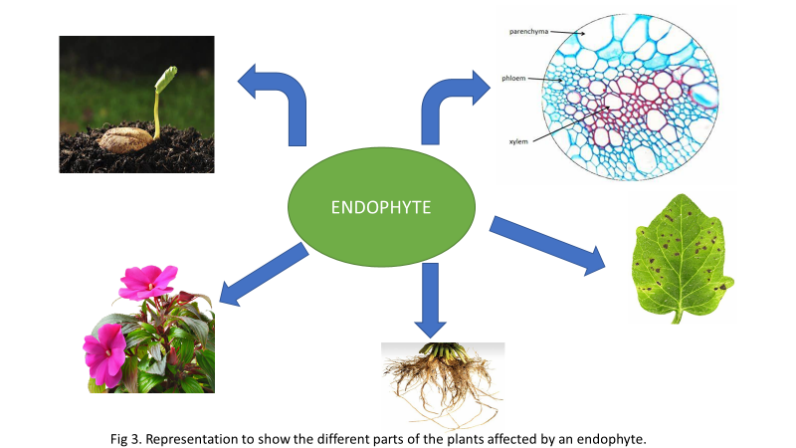
Figure 4: Representation to Show the Different Parts of the Plants Affected by an Endophyte
It has been stated that the foliar endophytic fungi in fallen leaves switch to a saprotrophic mode and help in litter decomposition which is one of the advantage of foliar endophytes on detritus cleaning [14]. Isolation of endophytes from various plant parts showed greater number of endophytes during monsoon than winter [15].
A number of medicinal plants have been found to be associated with endophytes. According to Sagar et al. [16], Eighteen Endophytic bacterial species were isolated from the different plant parts of Bryophyllum pinnatum where their abundance has been found to vary according to the season. Andrographis panniculata also known as ‘Kalmegh of Ayurveda’ has been found to be associated with twenty bacterial endophytes [17]. Another medicinal plant of importance is Ocimum sanctum from where 90 fungal endophytic species have been isolated where the abundance of the species is found to be affected by the mean temperature of the locality at the time of sampling [18]. It has been stated that the occurrence of the fungal endophytes is influenced by the type of host tissue and chemicals present in the medicinal herbs [19]. Fungal endophytes belonging to Ascomycetes have also been found to colonize the lichen thalli and the colonization rate was measured at 61.5 to 90.6% [20]. Histological investigation of the tissues can be used to study endophytic growth patterns, inter and intracellular nature, endophyte density in tissue, identify the part of tissue in which the endophyte is more concentrated [21].
Techniques Used for Identification of Endophytes
The study of endophytes is a challenging task as some of the endophytes may go unobserved in plants either due to their abundance or due to some other reasons. But in the last 10-15 years of most of the research studies based on endophytes, other than using the classical methods of identification, molecular approach is also being used. The advantage of using molecular approach is that even the sterile and unculturable endophytic species can be studied which is not possible in traditional approach. According to Tejesvi et al. [22], recent advances in DNA sequencing technology and computation allows more complete description of endophytic communities. For their proper identification, the endophytes need be isolated from the plant parts under study. The isolation methods are more or less same for all kinds of endophytic microbes, including fungi and bacteria [21].
There are two techniques by which endophytes can be studied- Classical or Traditional technique and Molecular technique. Traditional technique includes- direct observation method and cultivation dependent method. But non-sporulating and uncultivable endophytes molecular approach is used. Therefore, molecular technique comes into role as far as identification and study of endophytes are concerned. According to Paulo et al. by using Culture techniques only a small fraction of the bacteria diversity can be assessed because only few endophytes can be cultivated using laboratory methods hence molecular approach is used for the uncultivable endophytes. Be it the endophytic fungi, bacteria or the actinomycetes a dual approach consisting of cultivation and molecular approach is used for their identification. The basic mechanism involved in both the traditional method and the molecular method is depicted in Figure 5.
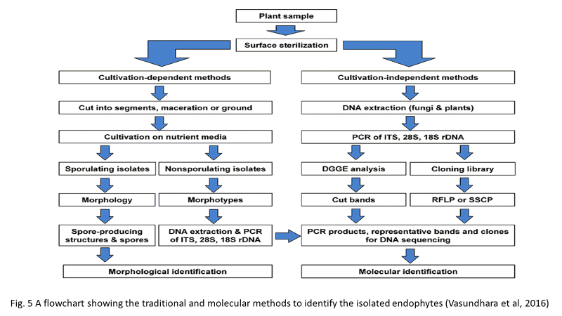
Figure 5: A Flowchart Showing the Traditional and Molecular Methods to Identify the Isolated Endophytes [33]
Classical Methods of Identification
For preliminary identification of the endophytes classical methods are used. These methods are based on morphological and biochemical tests that the endophytes respond to. Various staining methods, Culture plate methods are used for the isolation as well as the identification of the endophytes. The basic procedure involved has been found to be almost same in the different types of endophytes studied till date.
For the endophytic fungi morphological characteristics like sporulating structure were considered as diagnostic features for the morphological identification of endophytic fungi [23,24]. According to Sandhu et al. [25], Endophytic fungi are identified on the basis of the morphological characters which according to the standard taxonomic keys include colony diameter, texture, colour and the dimensions and morphology of hyphae and conidia. For morphological identification the spores of endophytic fungi are stained with lactophenol cotton blue staining. This stain imparts blue colour to the fungal hyphae and is therefore used for their identification. Apart from colony morphology classical identification of the endophytic fungi also involves observing the Shape and size of the spores, their growth and the rate of sporulation [26,27]. Apart from spore morphology their asexual and sexual state is also used for morphological identification of the endophytes [14].
Identification of Actinomycetes is done on the basis of cultural characteristics and morphology of the fruiting bodies and spores. Various characters include presence of conidial mycelium, spore mass colour, distinctive reverse colony colour, sporophore and spore chain morphology [28]. Presence of aerial mycelia, colour of diffusible pigments are also used for the identification of the Actinomycetes.
Visual observation of both morphological and Gram stain properties are also used for identification of Actinomycetes. In one of the study microscopic observation reveal that some of the endophytic Actinomycetes appear as dry, fuzzy, sticky, hard filamentous colonies. Chain morphology was studied under Field Emission Gun Screening Electron Microscopy [10].
The methods applied in carrying out the identification of the bacterial endophytes by the traditional methods include morphological and biochemical tests. Morphologically they are identified by observing the colonies formed, endospore formation and on the basis of the response to the Gram stain [29]. Biochemical tests include citrate utilization, Oxidase metabolism, nitrate reduction, Gelatin liquefaction, Starch hydrolysis, Catalase metabolism etc. Carbohydrate metabolism test is also done using sucrose, fructose, xylose, mannitol etc for the identification purpose. Shukla et al. [29], used Bergey’s Manual of Systemic Bacteriology to confirm the isolates identified. It is the main source for determining the identity of the bacterial species using every characterizing aspect. Structural and functional attributes are taken for classifying the organisms.
Molecular Methods of Identification
Traditional techniques used for the identification of the endophytes are very much common in all the research work dealing with the diversity studies of the endophytes. But it may not give the complete information as far as correct grouping of the endophytic species is concerned. In the past decade apart from the use of microscopy, staining and culture methods, standard techniques at the molecular level has also been used for the identification of the endophytic species. Molecular approaches has been widely used by the research workers in carrying out the different aspects of endophytic studies. Be it the diversity study or the one related to the bioprospecting of endophytes in most of the recent studies a combination of traditional and molecular methods has been used for the identification of the endophytic species in the host plant. The molecular approach is concerned with the genomic studies which is of great help in assessing the uncultivable bacterial as well as the fungal endophytic species.
The most commonly sequenced DNA regions are the ITS regions also called as Internal transcribed regions. It is used due to the high copy of the rRNA clusters and it is easy to detect even from small quantities of DNA. In most of the endophytic studies concerning Bacteria and Actinomycetes ITS 1 regions were studied which are associated to 16srRNA genes [10,30]. For the endophytic fungi ITS 1 associated with 18s and 5.8s rRNA genes and ITS2 associated with 5.8s and 28s rRNA genes have been sequenced [23]. Carolina M et al. 2014 has stated the use of 16s rRNA based techniques along with the use of T- RFLP and DGGE together to identify the endophytic bacteria at the molecular level. Verma et al. [21], reported the use of ITS 1, ITS 2 and ITS 4 as the frequently used primers for endophytic fungal identification. It has been reported in their study that as 16s rRNA has limited variability in some groups and limited representation in some databases hence it is often difficult to identify species in some bacterial groups. Gupta R et al. [31], has also mentioned the partial sequencing of 16srRNA genes and later comparing the gene sequences by BLAST (Basic Local Alignment Search Tool) technique. Many researchers are using this tool to identify the endophytic isolates.
The different techniques used for molecular studies include DNA fingerprinting, Polymerase Chain Reaction (PCR), Sequencing method, DNA barcoding method, Molecular phylogenetics, Biosensors, Immunosorbent assay etc.
The procedure involved in the molecular study is shown in the Figure 6.
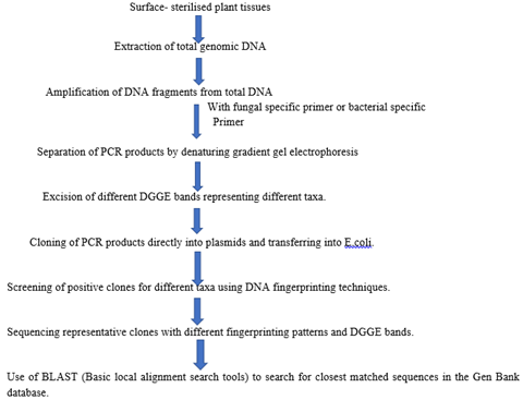
Figure 6: Flowchart Show the Procedure Involved in the Molecular Study
The different techniques used for molecular studies include DNA fingerprinting, Polymerase Chain Reaction (PCR), Sequencing method, DNA barcoding method, Molecular phylogenetics, Biosensors, Immunosorbent assay etc.
Some advanced genomic applications to study the endophytic species include using phenotype microarrays and analysis of the transcriptomes of both the host and the microbes, Suppression Subtractive Hybridization (SSH) technology and quantitative real time PCR [22]. Fungal specific primers ITS 4 and ITS 5 have been used for studying the distribution and diversity of the foliar endophytic fungi in the mangroves of the Andaman Islands [14].
In yet another study conducted on the bacterial endophytes it has been mentioned that Genomic studies for microbial communities include metagenomics, meta-proteomics and meta-transcriptomic [32]. Metagenomics not only focuses on a single gene marker but rather it gives importance to the sequencing the genomes of all the organism.
Genomes of filamentous fungi have been found to contain more gene clusters for secondary metabolite biosynthesis and hence selection of the appropriate genes or enzymes as markers for screening of the endophytic isolates is important [33]. According to Verma K et al [21], diversity of the endophytes can be studied by applying many methods like colonisation frequency, Isolation frequency, alpha diversity indices etc. Tejesvi et al. [22], has reported about the genomics application to study endophytes.
Discussion:
Endophytes have been reported from the different kinds of plants. Be it the medicinal plant like Bryophyllum [16] or Cereals like Oryza sativa [28], endophytes have been isolated from almost all kinds of plants. Diverse kinds of endophytes have also been isolated from the mangroves of the Andaman Islands where statistical analyses revealed the increase in the number of endophyte isolates with increase in the sample size [14]. By using both the classical methods and the molecular methods of identification, endophytes have been reported from the diverse kind of host plants.
Some of the common endophytes isolated from the different groups of plants has been shown in Table 1.
Table 1: List of Commonly Occurring Endophytes Studied in India
|
Category |
Name of the Species |
Host Plant |
Reference |
|
A. Fungi |
Alternaria alternate |
Bryophyllum pinnatum Lam. |
Sagar et al. [16]
|
|
Aspergillus niger |
|||
|
Fusarium moniliformi |
|||
|
Penicillium chrsogenum |
|||
|
Phoma sp. |
|||
|
Rhizopus sp |
|||
|
Aspergillus flavus |
Oryza sativa |
Naik et al. [28] |
|
|
Fusarium oxysporum |
|||
|
Penicillium chrsogenum |
|||
|
Trichoderma viridae |
|||
|
Chaetomiun globosum |
|||
|
Cladosporium cladosporoides |
|||
|
Alternaria raphanin |
Vitex negundo L |
Desale et al. [23] |
|
|
Aspergilus flavus gr |
|||
|
Colletotrichum gloeospoirides |
|||
|
Fusarium solani |
Calotropis procera |
Rani R et al. [35] |
|
|
Aspergillus nidulans |
|||
|
Aspergillus orzae |
|||
|
Chaetomium atrobrunemm |
|||
|
Aspergillus fumigatus |
Riccinus communis |
Sandhu et al. [25] |
|
|
Aspergillus japonicus |
|||
|
Fusarium semitactum |
|||
|
Curvalaria pallaescens |
|||
|
Fusarium solani |
|||
|
Aspergillus repens |
|||
|
Seridium abietinum |
Ocimum basalicum |
P. Palanichamy et al. [36] |
|
|
Mycelia sterilia |
|||
|
B. Actinomycetes |
Streptomyces albosporus |
Aloe vera |
Gangwar et al. [11] |
|
Streptococcus aureus |
Ocimum sanctum |
||
|
Streptococcus griesofuscus |
Mentha arvensis |
||
|
Actinopolyspora sp |
Ocimum sanctum |
||
|
Saccharopolyspora sp. |
Aloe vera |
||
|
Saccharopolyspora sp. |
Ocimum sanctum |
||
|
Saccharopolyspora sp. |
Mentha arvensis |
||
|
Streptomyces sp |
Mikania micrantha |
Momin and Tripathi [10] |
|
|
Nocardiopsis sp |
Costus speciosus |
||
|
Actinobacteria bacterium |
|||
|
C. Bacteria |
Rhizobium sp |
Oryza spp |
Banik et al. [37] |
|
Azospirillum sp |
|||
|
Rhizobium sp |
|||
|
Azotobacter sp. |
|||
|
Klebsiella spp |
|||
|
Actinobacteria sp |
|||
|
Bacillus subtilis |
Prosopis cineraria |
Gupta et al. [31] |
|
|
Stenotrophomus maltophilia |
|||
|
Bacillus spp |
Legume plants- Red gram, Black gram, Green gram, Cowpea and Chickpea |
Maheswari et al. [13] |
|
|
Micrococcus sp |
|||
|
Pseudomonas spp |
|||
|
Flavobacterium spp |
|||
|
Serratia sp |
|||
|
Bacillus sp |
Oryza spp |
Banik et al. [37] |
|
|
Ancylobacter sp |
|||
|
Klebsiella spp |
|||
|
Azotobacter spp |
|||
|
Aeromonas spp |
|||
|
Pseudomonas sp |
Different crops |
Vetrivelkalai et al. [38] |
|
|
Bacillus sp |
|||
|
Methylobacterium sp. |
A large number of Fungal endophytes have been isolated from the different group of plants under taken for the diversity study. But the isolation of Bacterial endophytes and that belonging to the Actinomycetes group is comparatively less in number. The difference in this aspect can be attributed to the type of interaction that exists between the host plant and the microbial species. Other than this interaction, other factors like the temperature, surrounding, climate of the area where the plants are there may affect the endophytic diversity. As the bacterial population is more than the fungal ones hence more extensive research is required for their presence in the plants. It is important to know the type of all the microbial forms found in a host plant so as to know the mechanics of the host plant. Another important fact is that the isolation of fungal species is easier as compared to the bacterial forms through culture method hence in most of the research work studied so far fungal endophytes have been reported in most of them as listed in Table 1. Therefore, apart from the application of the classical techniques efforts are being made for the use of modern tools and techniques for the identification of the endophytic species.
Roots, stems, petioles, leaves, lamina, inflorescence, rhizome exhibited a host of endophytes as reported in Table 3. The type of endophytes isolated range from Bacteria, Fungi to Actionomycetes. The characters used for identification of the isolated endophytic species revealed the use of both morphological and cultural characteristic features (Table 2).
Table 2: Morphological Characteristics Used for Identifying the Type of Endophytes
|
Category |
Morphological characters used for identification |
Reference |
|
Fungi |
Cultural characters, Morphology of fruiting bodies and spores |
Naik et al. [28] |
|
Sporulating structures |
Desale et al. [23] |
|
|
Conidial characters, Colony colour, Chlamydospore size, position and space |
Baruah et al. [39] |
|
|
Colony diameter, texture, colour and the dimension and morphology of hyphae and conidia |
Sandhu et al. [25] |
|
|
Morphology of the spores by using lactophenol cotton blue staining |
Palanichamy P et al. [36] |
|
|
Morphology of the surface texture, pigmentation, spores at the hyphal tips |
Chaturdevi and Gowrie, [40] |
|
|
By observing pycnidia, conidia and conidiogenous cells, conidiophores |
Suradkar et al. [41] |
|
|
Spore size, growth rate, sporulation rate, colony morphology, conidial shape and size |
Rabha et al. [26] |
|
|
Colony morphology, growth rate, hyphal mat characteristics, pigmentation of the fungal colony and medium. |
Nayak B K, [42] |
|
|
Bacteria |
Colony morphology, shapes, colour, margin, texture, Gram reaction, Endospore staining, motility |
Anjum N and Chandra R [43] |
|
Colony morphology according to Bergey’s manual of determinative Bacteriology |
Kumar A et al. [44] |
|
|
Gram staining and endospore formation |
Shukla et al. [29] |
|
|
Gram reaction, Endospore staining, Capsule staining, motility |
Arunachalam et al. [17] |
|
|
Actinomycete |
Conidial mycelium, spore mass colour, reverse colony colour, diffusible pigment, sporophore and spore chain morphology |
Naik et al. [28] |
|
Appearance of filamentous colony |
Momin and Tripathi, [10] |
|
|
Presence of aerial mycelia, spore mass colour, distinctive reverse colony colour, colour of diffusible pigments and spore chain morphology |
Gangwar et al. [11] |
Use of the classical methods for identification is applicable to the endophytic species which are culturable but for the ones which remains uncultured are labelled as sterile mycelia. Molecular techniques have found wide application in identifying the ones which cannot be studied by using the classical or traditional methods of identification. According to Paulo et al. only a small fraction of the bacterial diversity can be assessed by using isolation methods because only few can be cultivated using traditional laboratory methods. The number of endophytes isolated from the different host plants represents variation in the number of the endophytes isolated (Table 3).
Table 3: List of the Number of Endophytes Isolated from the Host Plant
|
Host Plant |
Endophyte type |
No. of Endophytes |
Reference |
|
Bryophyllum pinnatum Lam. |
Fungi |
18 |
Sagar A et al. [16] |
|
Oryza sativa |
Fungi |
19 |
Naik et al. [28] |
|
|
Actinomycete |
01 |
Naik et al. [28] |
|
Andrographis paniculata |
Bacteria |
20 |
Arunachalam et al. [17] |
|
Vitex negundo |
Fungi |
17 |
Desale et al. [23] |
|
Ocimum sanctum |
Fungi |
40 |
Pavithra N et al. [24] |
|
Calotropis procera |
Fungi |
20 |
Nirwaan et al. 2017 |
|
Riccinus communis |
Fungi |
10 |
Sandhu et al. [25] |
|
Justicia gendarusa |
Fungi |
05 |
Palanichamy P et al. [36] |
|
Madhuea longifolia |
Fungi |
03 |
|
|
Vitex negundo |
Fungi |
06 |
|
|
Flacourtia jangomas |
Bacteria |
13 |
Shukla et al. [29] |
|
Embilica officinalis |
Bacteria |
04 |
|
|
Catharanthus roseus |
Bacteria |
02 |
|
|
Cissus quandrangularis |
Fungi |
05 |
Chathurdevi and Gowrie, [40] |
|
Cissus quadrangularis |
Fungi |
08 |
Suradkar et al. [41] |
|
Camellia sinensis |
Fungi |
30 |
Rabha et al. [26] |
|
Pongamia pinnata |
Fungi |
15 |
Nayak B K [42] |
|
Ocimum sanctum |
Fungi |
90 |
Chowdhary and Kaushik [18] |
|
Aloe vera |
Actinomycete |
06 |
Gangwar et al. [11] |
|
Mentha arvensis |
Actinomycete |
18 |
|
|
Ocimum sanctum |
Actinomycete |
16 |
|
|
Mikania micrantha |
Actinomycete |
07 |
Momin and Tripathi [10] |
|
Ageratum conizoides |
Actinomycete |
02 |
|
|
Costus speciosus |
Actinomycete |
02 |
|
|
Cassia fistula |
Actinomycete |
02 |
|
|
Scopariadulcis |
Actinomycete |
02 |
|
|
Curcuma longa |
Bacteria |
14 |
Kumar A et al. [44] |
|
Catharnthus roseus |
Bacteria |
35 |
Anjum N and Chandra R, [43] |
|
Ocimum sanctum |
|||
|
Mentha arvensis |
|||
|
Stevia rebaudiana |
As stated above this difference in the number of endophytes isolated can be attributed to the overall conditions in which the host plant is found to be grown. Naik et al [28], were successful in isolating both Fungal and Actinomycetes from Oryza sativa.
Endophytes have been isolated from a number of plants specially the medicinal plants. Research work has shown that the medicinal plants secrete bioactive substances that are of pharmacological importance [34]. Endophytic fungi isolated from Riccinus communis exhibits a great antibacterial activity against six human pathogenic bacteria [25]. Studies done on Bryophyllum pinnatum by Sagar et al [16], showed maximum antibacterial activity against Staphylococcus aureus at all concentrations as compared to E. coli and Y. pestis. The benefits of the medicinal plants have an association with the endophytic species found within them. But the specificity of the endophytic species with the type of bioactive substance released by the plants needs extensive research which will be a time taking process. According to Vasundhara M et al. [33], out of the total endophytic fungi harbouring each plant species only a minor fraction of them are able to produce important metabolites and to identify the potential isolate capable of producing a particular compound one needs to screen all isolated endophytes that runs into hundreds. This is also one of the reason for the research workers to isolate a specific group of endophytes for conducting diversity studies.
In the last decade much research has been conducted in the field of Endophytes and the boom in this field of research around the world is throwing light on the future development that will take place in the use of bio products in pharmacology. Many medicinal plants apart from cereals and other plants are being researched for the presence of endophytes within them and their role as antifungal or antibacterial agents. But less is being done in finding out the type and nature of the specific endophytic species responsible for the antimicrobial property. In India as well the North Eastern part of India more extensive research is required which may help the pharmacological sector in India.
As per Rabha et al. [26], more studies are required to analyse the genetic variation among the isolates of a species with different markers and thereby to establish a proper relationship between the morphological and genetic variation.
A lot of research has been going on to unravel and exploit the advantages that the endophytes can have in human welfare. Scientists have been successful in unravelling the fact behind the bioactive potential of the endophytes and its importance in the agricultural as well as pharmacological sector. Research in the field of endophytes have shown that they provide a wide range of benefits to the host plant. Endophytic species like Bacteria, Fungi as well as Actinomycetes have been found to promote production of growth promoting substances like Indole acetic acid, IAA [13]. Other than having growth promoting activity endophytes have been found to be potent as antimicrobial agents. They have been found to be having antibacterial and antifungal activity [11,16,35]. Numerous research work in the field of bioactivity in the Medicinal plants of India has taken place where they been found to be having anti-microbial activity as well as potent biocontrol agent [18].
CONCLUSIONS
To understand the mechanics of endophytic activity in the host plant, variety of research work has been conducted. Right from diversity studies to their activity as biocontrol agent as well as knowing about their biofuel activity a number of active work has been conducted and the more active research needs to be done focussing more on the genetics of endophytes and their utility in extracting bioactive agents from the medicinal plants. By applying modern tools and advanced techniques more information related to endophytic relationship with the host can be worked out.
This review has been done to bring out the different aspects of endophytic diversity and to elaborate on the different tools to identify the endophytic isolates.
A number of work has been carried out in the last 10-15 years on endophytes in diverse aspects all over the world including India but the number of research work focusing more on diversity aspect is quite less. North Eastern part of India being one of the biodiversity rich part of it needs more focus on the endophytic research as from this part of the country work on endophytic diversity is scarce. Studies on endophytic diversity with the use of modern techniques will help a lot in unravelling the hidden aspects of the microbial world in the field of their bio prospects.
Acknowledgement
Authors are thankful to ‘The Assam Royal Global University’ for providing the structural infrastructure for carrying out the study.
REFERENCES
- Stone, J.K., et al. “An overview of endophytic microbes: endophytism defined.” Microbial Endophytes, edited by C.W. Bacon and J.F. White, M Dekker Inc, 2000, pp. 3–5.
- Suryanarayanan, T.S., et al. “The host range of multi-host endophytic fungi.” Current Science, vol. 115, no. 10, 2018, pp. 1963–1969.
- Kandel, S., et al. “Bacterial endophyte colonization and distribution within plants.” Microorganisms, vol. 5, 2017, pp. 1–27.
- Ahmad, R., et al. “A brief history of endophyte detection techniques in grasses.” Sustainable Agriculture Research, vol. 8, no. 3, 2019.
- Schulz, B., et al. “What are endophytes?” Soil Biology, vol. 9, 2005.
- Kogel, K.H., et al. “Endophyte or parasite: What decides?” Curr Opin Plant Biol, vol. 9, 2006, pp. 358–363. https://doi.org/10.1016/j.pbi.2006.05.001.
- Bernard, S., et al. “Botryosphaeriaceae as endophytes and latent pathogens of woody plants: Diversity, ecology and impact.” Fungal Biology Reviews, vol. 21, 2007, pp. 90–106.
- Rodriguez, R.J., et al. “Fungal endophytes: Diversity and functional roles.” New Phytologist, 2009, pp. 1–9. https://doi.org/10.1111/j.1469-8137.2009.02773.
- Bayman, P. and T.J. Otero. “Microbial endophytes of orchid roots.” Soil Microbiology, vol. 9, 2006, pp. 153–181.
- Momin, M.D. and S.K. Tripathi. “Studies of endophytic actinomycetes associated with medicinal plants of Mizoram, northeast, India.” International Journal of Current Microbiology and Applied Sciences, vol. 7, no. 12, 2018, pp. 1398–1407.
- Gangwar, M., et al. “Diversity and biopotential of endophytic actinomycetes from three medicinal plants in India.” African Journal of Microbiology Research, vol. 8, no. 2, 2014, pp. 184–191.
- Kumar, A., et al. Microbial endophytes: Functional biology and applications. 2019.
- Maheswari, T.U., et al. “Studies on phytohormone producing ability of indigenous endophytic bacteria isolated from tropical legume crops.” International Journal of Current Microbiology and Applied Sciences, vol. 2, no. 6, 2013, pp. 127–136.
- Rajamani, T., et al. “Distribution and diversity of foliar endophytic fungi in the mangroves of Andaman Islands, India.” Fungal Ecology, vol. 36, 2018, pp. 109–116.
- Nalini, M.S., et al. “Endophytic fungal diversity in medicinal plants of western ghats, India.” International Journal of Biodiversity, 2014, pp. 1–9.
- Sagar, A., et al. “Studies on endophytes and antibacterial activity of bryophyllum pinnatum Lam.” International Journal of Applied Research, vol. 3, no. 1, 2017, pp. 126–134.
- Arunachalam, C. and P. Gayathri. “Studies on bioprospecting of endophytic bacteria from the medicinal plant andrographis paniculata for their antimicrobial activity and antibiotic susceptibility pattern.” International Journal of Current Pharmaceutical Research, vol. 2, no. 4, 2010, pp. 63–64.
- Chowdhary, K. and K. Nutan. “Fungal endophyte diversity and bioactivity in the indian medicinal plant ocimum sanctum linn.” PLoS One, vol. 10, no. 11, 2015.
- Rajagopal, K., et al. “Diversity of fungal endophytes in few medicinal herbs of south India.” Asiana Journal of Experimental Biological Sciences, vol. 1, no. 2, 2010, pp. 415–418.
- Vinayaka, K.S., et al. “Association and variation of endophytic fungi among some macrolichens in central Western Ghats, Southern India.” Int J Curr Microbiol App Sci, vol. 5, no. 6, 2016, pp. 115–124.
- Verma, S., et al. “Exploring Endophytic communities of plants: methods for assessing diversity, effects on host development and potential biotechnological applications.”
- Tejesvi, M.V., et al. “Phylogenetic analysis of endophytic pestalotiopsis species from ethnopharmaceutically important medicinal trees.” Fungal Diversity, vol. 38, 2009, pp. 167–183.
- Desale, M. and M.G. Bodhankar. “Antimicrobial activity of endophytic fungi isolated from vitex negundo linn.” Int J Curr Microbiol App Sci, vol. 2, no. 12, 2013, pp. 389–395.
- Pavithra, N., et al. “Antimicrobial and enzyme activity of endophytic fungi isolated from tulsi.” Journal of Pharmaceutical and Biomedical Sciences, vol. 16, no. 12, 2012.
- Sandhu, S.S., et al. “Isolation and identification of endophytic fungi from ricinus communis linn and their antibacterial activity.” IJRPC, vol. 4, no. 3, 2014, pp. 611–618.
- Rabha, A., et al. “Morphological and molecular diversity of endophytic colletotrichum gloeosporioides from tea plant, camellia sinensis (L.) O. kuntze of assam, India.” Journal of Genetic Engineering and Biotechnology, vol. 14, 2016, pp. 181–187.
- Rana, L.K., et al. “Biodiversity of endophytic fungi from diverse niches and their biotechnological applications.” Advances in Endophytic Fungal Research, 2019. https://doi.org/10.1007/978-3-030-03589-1_6.
- Naik, S.B., et al. “Study on the diversity of endophytic communities from rice (Oryza Sativa L.) and their antagonistic activities in vitro.” Microbial Research, vol. 164, 2009, pp. 290–296.
- Shukla, S., et al. “Potential antimicrobial activity of bacterial endophytes isolated from Flacourtia Jangomas (Lour) raeusch, a less explored medicinal plant.” J Microbiol Biotech Food Sci, vol. 4, no. 6, 2015, pp. 473–477.
- Lata, K.R., et al. “Endophytic microbiomes: Biodiversity, ecological significance and biotechnological applications.” Research Journal of Biotechnology, vol. 14, no. 10, 2019.
- Gupta, R.M., et al. “Isolation, characterization and identification of endophytic bacteria by 16S rRNA partial sequencing technique from roots and leaves of prosopis cineraria plant.” Asian Journal of Plant Science and Research, vol. 5, no. 6, 2015, pp. 36–43.
- Maela, P. and M. Dlamini. “Current understanding of bacterial endophytes, their diversity, colonization and their roles in promoting plant growth.” Applied Microbiology, vol. 5, no. 1, 2019, pp. 1–12.
- Vasundhara, M., et al. “Molecular approaches to screen bioactive compounds from endophytic fungi.” Frontiers in Microbiology, vol. 7, 2016, pp. 1774.
- Quecine, M.C., et al. “Diversity and biotechnological potential of plant-associated endophytic bacteria.” Plant Biotechnology, vol. 2, 2014, pp. 377–423.
- Rani, R., et al. “Antibacterial activity of twenty different endophytic fungi isolated from Calotropis Procera and time kill assay.” Clinical Microbiology, vol. 6, 2017, pp. 280.
- Palanichamy, P., et al. “Bioactive potential of secondary metabolites derived from medicinal plant endophytes.” Egyptian Journal of Basic and Applied Sciences, vol. 5, 2018, pp. 303–312.
- Banik, A., et al. “Characterization of N₂-fixing plant growth promoting endophytic and epiphytic bacterial community of indian cultivated and wild rice (Oryza spp.) genotypes.” Planta, vol. 243, 2016, pp. 799–812.
- Vetrivelkalai, P., et al. “Biocontrol potential of endophytic bacteria on meloidogyne incognita and its effect on plant growth in bhendi.” Journal of Biopesticides, vol. 3, no. 2, 2010, pp. 452–457.
- Baruah, N., et al. “In vitro screening of native banana rhizospheric microbes and endophytes of assam against Fusarium Oxysporum f. sp. Cubense.” Int J Curr Microbiol App Sci, vol. 7, no. 6, 2018, pp. 1575–1583.
- Chathurdevi, G. and S. Gowrie. “A study on the bioactive potential of endophytic fungi isolated from medicinal Plant.” European Journal of Biomedical and Pharmaceutical Sciences, vol. 2, no. 4, 2015, pp. 848–866.
- Suradkar, K.P. and D.V. Hande. “Morphotaxonomy of endophytic fungi on cissus quadrangularis from amravati (MS), India.” Journal of Bacteriology and Mycology, vol. 5, no. 2, 2017, pp. 253–258.
- Nayak, B. “Studies on endophytic fungal diversity from different leaf samples of pongamia pinnata.” International Journal of MediPharm Research, vol. 1, no. 2, 2015, pp. 134–138.
- Anjum, N. and R. Chandra. “Endophytic bacteria: optimization of isolation procedure from various medicinal plants and their preliminary characterization.” Asian J Pharm Clin Res, vol. 8, no. 4, 2014, pp. 233–238.
- Kumar, A., et al. “Isolation and characterization of bacterial endophytes of curcuma longa L.” 3 Biotech, vol. 6, 2016, pp. 60.